Arterial and Venous Ultrasound
Arterial and Venous Ultrasound

What is Arterial and Venous Ultrasound?
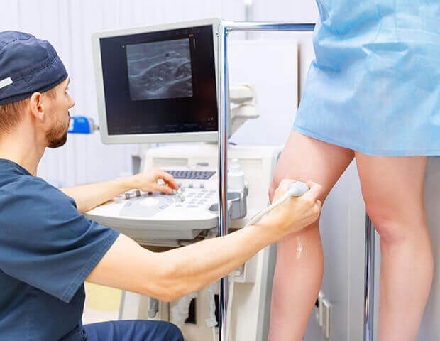
Arterial and Venous Ultrasound uses high-frequency sound waves to visualize the body’s arterial and venous systems and assess blood flow. A handheld device called a transducer is moved over the area as sound waves bounce off blood cells, creating images of vessels on a monitor.
An important use of venous ultrasound is creating detailed maps of the veins, known as vein mapping. This technique utilizes ultrasound technology to reveal the size, depth, and blood flow of the veins, providing a non-invasive method for assessment. This information is essential for effectively planning treatments for conditions like superficial venous reflux and varicose veins.
Risks and Benefits of Arterial and Venous Ultrasound
Arterial and venous ultrasounds offer numerous benefits that outweigh the minimal associated risks. These procedures provide valuable insights into vascular health, facilitating accurate diagnoses, early detection, and personalized treatment planning. If you have concerns about your cardiovascular health or have risk factors for cardiovascular conditions, consulting a cardiologist and undergoing an arterial and venous ultrasound can play a pivotal role in maintaining your cardiovascular well-being.
Benefits of Arterial and Venous Ultrasound
- Non-Invasive
Arterial and venous ultrasounds are non-invasive and painless procedures. They do not involve radiation exposure, making them safe for patients of all ages. - Accurate Diagnosis
Ultrasounds provide real-time images of blood vessels, allowing accurate visualization of blood flow, blockages, and abnormalities. This aids in the early detection and diagnosis of various vascular conditions. - Early Detection
Vascular ultrasounds can detect conditions like arterial stenosis, blood clots, aneurysms, and venous insufficiency at an early stage. Early detection allows for timely intervention and better treatment outcomes. - Tailored Treatment Plans
With detailed images of the blood vessels, vascular specialists can create personalized treatment plans that address specific patient needs. This precision leads to more effective and targeted interventions. - Monitoring Progress
Vascular ultrasounds enable ongoing monitoring of vascular conditions, helping doctors track changes over time and adjust treatment strategies as necessary.
Risks include but are not limited to:
- Minimal discomfort
- Rare allergic reactions
- Limited imaging depth
What to Expect Before, During, and After an Arterial and Venous Ultrasound
Before undergoing an arterial and venous ultrasound, there's generally minimal preparation needed. It is advised that you drink at least four (4) cups of water the morning of the ultrasound.
It is also important that you do NOT wear compression stockings 24 hours prior to the scheduled cardiology test and procedures. Wearing the compression stockings within 24 hours prior may invalidate the test results, causing the test to be rescheduled.The test takes approximately forty-five (45) minutes. Be prepared to undress from the hip down.
During the procedure, you will be asked to lie down on an examination table. A water-based gel will be applied to the area being examined, ensuring good contact between your skin and the transducer. The vascular technologist will move the transducer over the skin in the region of interest to capture images from various angles. This process is painless and non-invasive, and you might hear gentle whooshing sounds as the sound waves interact with the blood flow. After the arterial and venous ultrasound, you can immediately resume your regular activities. Here are a few things to keep in mind post-ultrasound:
- Depending on the results, further diagnostic tests or treatment options might be recommended.
- Your specialist will discuss the findings with you, address any concerns, and collaborate on a personalized care plan.
- If you have known vascular conditions, regular ultrasounds could be advised to monitor changes over time.
Am I a Candidate for Arterial and Venous Ultrasound?
Early detection and proactive management of vascular conditions can lead to better outcomes and improved quality of life. If you suspect you may benefit from arterial and venous ultrasound, you should talk to your cardiovascular specialist. If you are experiencing symptoms or have risk factors associated with vascular conditions, you may be a suitable candidate for this imaging technique. Here are some situations where arterial and venous ultrasound might be recommended:
- Peripheral artery disease (PAD) symptoms
If you are experiencing symptoms such as leg pain, cramping, numbness, or weakness, especially during physical activity, you may have peripheral artery disease. Arterial ultrasound can help assess blood flow in the arteries of your legs and arms, providing valuable information about potential blockages. - Varicose veins or venous insufficiency
If you have visible varicose veins, leg swelling, or pain, venous ultrasound can evaluate the condition of the veins close to the skin's surface. This procedure can help determine if you have venous insufficiency, a condition where veins have difficulty sending blood from the legs back to the heart. - Deep vein thrombosis (DVT) concerns
If you experience symptoms like leg pain, swelling, or warmth, it could be indicative of deep vein thrombosis (DVT). A venous ultrasound is instrumental in detecting blood clots in the veins. This information is crucial for prompt intervention and the prevention of complications. - Monitoring blood clot treatments
If you are undergoing treatment for blood clots, regular arterial and venous ultrasound can be used to monitor the effectiveness of the treatment and track any changes in the clot. - Vascular anomalies or conditions
If you have a known vascular anomaly, congenital condition, or require ongoing monitoring of your vascular health due to medical history or risk factors, arterial and venous ultrasound can provide valuable insights into your blood vessel structure and function.
Take a Closer Look at Your Vascular Health with Arterial and Venous Ultrasound
Photo Gallery
Video Gallery
Testimonials
Photo Gallery
Get To Know Our Cardiologists

